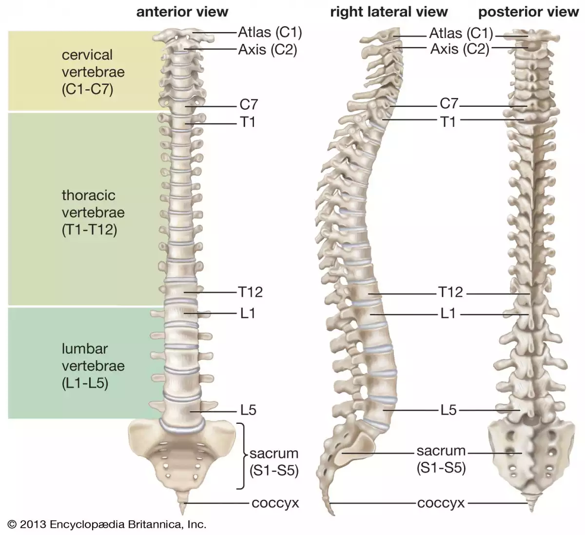
We will travel back to our formative years in primary school and reexamine the question: "How many vertebrae does the human body have?". Only this time around we will have a more in-depth look. The backbone is an essential element in the anatomy of all vertebrates and represents the basic criteria of classification.
How many vertebrae does our spine have?
The spinal column is the structure that allows movement, and in the case of humans, maintains us in an upright position. It is made out of small irregular bones called vertebrae that are aligned in an interlocking sequence from the base of the skull down to the pelvis. The backbone is divided into sections, each one with its specific vertebrae and functions. Therefore, discovering how many vertebrae we have also reveals their name and location within the body.
Vertebrae get their name depending on which area of the spinal column they are in (cervical, thoracic, lumbar, sacrum, and coccyx) along with a number, in ascending order, corresponding to its position in the given section of the backbone. For example, the 1st cervical vertebrae can be simply called "C1", and the same logic applies to the 4th vertebrae in the lumbar area, the 3rd in the sacrum or the 9th thoracic one, which will become L4, S3, and T9, respectively. In the case of coccygeal vertebrae, the abbreviation is "Co."
In total, the human body has 33 vertebrae which are placed in the following way: 7 cervical, 12 thoracic, 5 lumbar, 5 sacral, and 4 coccygeal vertebrae (the number is sometimes increased by an additional vertebra in one region, or it may be diminished in another). This mental math exercise may not render the same answer to everyone, as certain people don't include the sacrum and the coccyx. The argument is that once we reach adulthood, the two last sections fuse and become separate from the rest of the spinal column, however, for our investigation we will look at all the vertebrae as a whole.

1. Cervical vertebrae
There are seven cervical vertebrae designated C1 through C7, but only the first two get a special mention.
C1 or “Atlas”
The name references the Greek Titan that Zeus punished to stand on the edge of the world and hold up the sky on this shoulders. Therefore, the C1 vertebrae, much like the eponymous hero, supports the weight of the human head.
C2 or “Axis”
The Axis (from Latin axis = axle) forms the pivot upon which the Atlas rotates, and the structure of these two vertebrae is the reason why the neck and head have a broad range of motion. An interesting fact is that the C2 is only found in evolved animals such as reptiles, birds, and mammals (births and amphibians do not have this feature).
The C3 vertebra lies snugly underneath the skin and the other spinal bones and provides a passageway to nerve endings that irrigate the face, teeth, and ears. The following three vertebrae, C4, C5, and C6 have speech functions as they pass sound through the larynx and vocal cords which allow sound production. The C7 vertebra is distinctively long and is responsible for the articulation of our superior limbs.
2. Thoracic vertebrae
The thoracic vertebrae are unique among the bones of the spine in that they are the only vertebrae that support ribs and have overlapping spinous processes. Another vital function of the thoracic vertebrae is rotating the upper body.
Discomfort in any of the superior vertebrae, from T1 to T9 approximately, may cause breathing and heart problems. On the other hand, any interferences with the T9 - T12 vertebrae can cause digestive problems, allergic reactions, and pain in the lower limbs.
3. Lumbar vertebrae
The five lumbar vertebrae are the largest of the vertebrae, their robust structure being necessary for supporting more weight than the other vertebrae. Due to their flexing and extending qualities, they allow us to bend our torso forward as well as restrict the movement if we want to arch our back.
One of the defining characteristics of lumbar vertebrae is their curvature. Kidney shaped when viewed superiorly, wider from side to side than from front to back, and a little thicker in front than in the back. The outward curvature is present in all humans, but some people exhibit a more pronounced arch, which leads to a medical condition called lordosis.
Due to its proximity to the thoracic vertebrae which are involved in our digestive system, the L1 and L2 also play an important role in any gastrointestinal disease. The following vertebra, L3 deals with issues in the reproductive system while L4 incorporates the sciatic nerve. Pressure on the sciatic nerve causes pain that radiates from your lower back into the back or side or your legs.
4. Sacrum and coccyx
As a personal choice, many people leave out the sacrum and coccyx when they count the number of vertebrae in our spinal column. The reality is that we need to look at these two segments as being part of a whole and add them to the count as an integral part of our backbone.
The first three sacral vertebrae act as conductors for the reproductive system while the last two deal with complications associated with the urinary tract, stone formation, and bowel movement.
The coccyx, commonly referred to as the tailbone, is the final segment of the vertebral column in all apes and is the remnant of a vestigial tail.
References:
Encyclopedia Britannica, Vertebral column, https://www.britannica.com/science/vertebral-column

