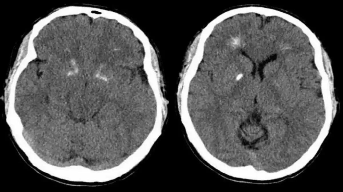
The human brain, weighing in at approximately 1.4 kg, is the body's largest and most complex organ. Inside the brain, certain structures keep the rest of the systems functioning in tip-top shape. This is why any brain injury or disease could cause the failure of any one of the body's essential functions.
The basal ganglia form one of these groups of cerebral nuclei. As a complex structure, the function of basal ganglia is still somewhat mysterious. That said, in this article, we'll give you a basal ganglia definition, and explain their anatomy and function with a diagram and a video.
Basal ganglia definition
The basal ganglia are a brain structure made up of a series of subcortical nuclei located at the base of the brain. This system of tiny nuclei interconnects with other brain structures like the cerebral cortex, thalamus, and other regions of the brain. Although new functions of this midbrain structure continue to be discovered, scientists know that they play a crucial role in operations such as controlling voluntary movements, learning processes, habits, working memory, eye movement, cognition, and emotions.
Based on various theories regarding their basic function, research suggests that these brain structures are mostly involved in one's ability to make decisions and to choose behaviors and the movements that we make at any given time. Each of the basal ganglia's parts has a complex anatomic structure and neurochemistry, making this one of the most complex and substantial systems both in the nervous system and in the whole human body in general.
Since this brain structure plays an important role in many of the body's functions, neurological conditions associated with some type of dysfunction in this structure stand out since they cause difficulties in the areas previously mentioned. Here are the main basal ganglia disorders:
Parkinson's Disease
Huntington's Disease
Tourette's Syndrome
Obsessive-compulsive disorder (OCD)
Dystonia
Addictions
Scientific evidence backs up the idea that the limbic part of the basal ganglia is closely connected to reward-based learning since dopamine is the main neurotransmitter in some of their pathways. So this explains the connection to addictions to certain drugs like nicotine, cocaine, and amphetamines.
Related: Nervous Tics: What They Are And How To Stop Tics Naturally
Basal ganglia anatomy
As previously mentioned, basal ganglia are fundamental brain structures that assemble different gray matter nuclei stored in the deepest regions of the brain. Anatomically speaking, this brain structure has four parts or distinct nuclei. Two of them: the striatum and the globus pallidus are the largest structures, while the substantia nigra and the subthalamic nucleus, are considerably smaller.

1. Striatum
The striatum is a subcortical structure, divided into the dorsal striatum and the ventral striatum. These nuclei are mainly formed by neurons from the GABAergic system that project towards other nuclei like the globus pallidus and the substantia nigra. Scientists believe that the dorsal striatum plays a fundamental role in sensorimotor activities, while the ventral side is related to reward-based learning, among other limbic functions. At the same time, this nucleus is made up of different structures including the caudate nucleus, the putamen, the nucleus accumbens, and the olfactory tubercle.
2. Globus Pallidus
The globus pallidus is considered a single neural mass. However, functionally, it can be divided into two sections, called the internal and external segments. Both sections contain mainly GABAergic neurons and participate in different neural circuits.
3. Substantia nigra
The substantia nigra is also a part of the basal ganglia. This substance is made up of gray matter and has two substructures: the pars compacta and the pars reticulata. While the pars reticulata works in a similar way to other structures like the globus pallidus, the pars compacta takes care of the dopamine production needed to maintain the equilibrium of the nigrostriatal pathway.
4. Subthalamic nucleus
Finally, the subthalamic nucleus is the only basal ganglia structure that produces an excitatory neurotransmitter: glutamate (other structures produce inhibitory neurotransmitters). The function of the subthalamic nucleus is to stimulate the activity of the pars reticulata and the globus pallidus.
Related: Neurotransmitters: Definition, 10 Main Types And Functions
Basal ganglia function
Throughout the article, we've described the diverse functions of the basal ganglia. Due to the complexity of these structures, currently, scientists continue to study the implications that this structure has on different body functions. However, we do know that this structure is essential to the following functions:
1. Eye movement
The role of the basal ganglia in controlling eye movement is one of the most studied functions of this structure. The super colliculus mainly controls this region of the brain. However, this area receives strong inhibitory projection from this structure.
2. Decision making
Studies propose two explanatory models regarding the implication of the basal ganglia in decision making. In one of them, a 'critical' agent, located in the ventral striatum, evaluates the possibility to take action and an 'acting' agent from the dorsal striatum carries out the action. On the other hand, the second model suggests that this brain structure functions as a selection mechanism. This means that the basal ganglia choose the actions that occur in the cortex based on the context.
Related: What Are Cells? Differences Between Prokaryotic And Eukaryotic Cells
3. Working memory
Various hypotheses on the relationship between the basal ganglia and working memory suggest that one of the functions of this structure is to regulate the working memory, controlling the information recorded within it and deleting the things that aren't.
4. Motivation
Research on animals, specifically rodents, suggests that the extracellular dopamine that travels through the basal ganglia has a close connection to the motivation and reward processes. Especially with the need to satisfy that sense of 'euphoria. In the following video, you'll find an explanation of this structure found in midbrain and its function:
2-Minute Neuroscience: Basal Ganglia
Check out the original article: ¿Qué son los ganglios basales? Partes y funciones at viviendolasalud.com
References
Chakravarthy, V. S., Joseph, D. & Bapi, R. S. (2010). What do the basal ganglia do? A modeling perspective. Biological Cybernetics, 103(3): 237–253.
Lanciego, J. L., Luquin, N. & Obeso, J. A. (2017). Functional Neuroanatomy of the Basal Ganglia. Cold Spring Harbor Perspectives in Medicine, 2(12).
