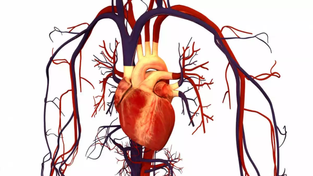
The heart is the human circulatory system's main organ. It's in charge of sending blood to tissue and organs all over the body. So, to learn more about its anatomy and physiology, in this article we explain all 21 parts of the heart and how each one functions. Besides, we tell you exactly where your heart is located, the tissues that it's made up of and how blood circulation works.
Parts of the heart and heart anatomy
Anatomy is the science that studies the body's shape and structure (both in humans and other animals). In other words, scientists in this field work to describe its parts. On the other hand, physiology is the science that describes the function of these body parts. The said, the heart is a hollow muscle with several different cavities, whose primary function is to permit blood flow.
The aforementioned occurs using two main movements: diastole, when the heart relaxes attracting blood to itself from the veins; and systole, when it contracts, releasing blood to the arteries. Muscle tissue known as myocardium (heart muscle), is the main component of this organ, which is broken down further into endocardium (the inner part) and epicardium (the external part). Besides, pericardium, a group of fibers that separates the heart from other organs, covers it. All of these are known as the layers of the heart.
Related: Muscles: What They Are, How Many We Have And Main Types
Where is your heart?
In the human body, the heart is located inside of the thorax, on the left-hand side (specifically inside of a compartment called the "mediastinum"). The lungs are located on either side of this muscle. The diaphragm is located below, and above there is an orifice called the "superior thoracic aperture." Also, the spinal cord is directly behind this organ. That's why we say that it has four sides: anterior (sternocostal), inferior (diaphragmatic), left pulmonary and right pulmonary. Its shape is comparable to a tilted cone.
Human circulatory system (cardiovascular system)
The heart forms a part the human circulatory system, also known as the cardiovascular system. This system is a group of organs and tissues whose function is to allow blood circulation. In other words, it's in charge of transporting blood inside of the body, and along with this, nutrients, oxygen, hormones, and other vital elements to different tissues. The cardiovascular system is mainly made up of the heart and blood vessels.
What is blood circulation?
In 1628, An English doctor named William Harvey discovered that blood follows a specific path inside the body: one of the parts of the heart called the "left ventricle" contracts pushing blood from the arteries to the aorta. From there, the blood spreads to the rest of our organs (besides the lungs). When it reaches different tissues, blood is collected by the veins and travels to the right atrium.
Then it travels towards the right ventricle, and from there it goes to the lungs. Inside of these respiratory organs, it oxygenates, and once this is complete, it returns to the left ventricle and left atrium, ending the cycle. This process is known as "blood circulation," and it plays a fundamental role in keeping the tissues alive and in the body of any animal in general.
Related: Circulatory System: What It Is, Parts And Functions
The 21 parts of the heart and their functions
The heart is a highly complex muscle, and there are many classifications to study it by. Generally speaking, the left and right sides of the heart are separated. Each side is made up of two cavities (four in total): the right atrium, left atrium (both located in the superior part of the heart), the right ventricle and the left ventricle. A septum, which has three parts, divides these parts of the heart.
So, based on several different human anatomy publications (Rutz and Pabst, 2007; Latarjet and Ruiz Liard, 2008; Palastanga, Field and Soames, 2000) we'll take a look at the 21 parts of the heart (divided into the right and left side and the septums that unite them).
Heart septums
This is the part of the heart that divides the right and left sides (this is its primary function). At the same time, the septum breaks down into three structures that have different thicknesses: the interatrial septum, interventricular septum, and the atrioventricular septum.
1. Interatrial septum
The interatrial septum is approximately 4 mm thick. At its center, the right atrium's fossa ovalis is found, which is what connects the two sides of the heart.
2. Interventricular septum
This septum's function is to separate the right and left ventricles. It is thicker than the interatrial septum and can be up to 12 mm wide, the closer it is to the cardiac muscle.
3. Atrioventricular septum
The atrioventricular septum is located between the two septums as mentioned earlier. This one connects the tricuspid and mitral valves. This is the thinnest heart septum.
Right heart
In anatomy and physiology, the right side of the heart is also called the "right heart." During the blood circulation cycle (which we will explain later on), the right heart's function is to receive blood. This is the general function of all of its parts in physiological terms, although anatomically speaking, each has a different role that helps to maintain the heart's structure. In large part, it is made up of two central cavities: an atrium and a ventricle, including veins, arteries, and valves. Below we take a look at their primary roles.
4. Right atrium
The right atrium is the right heart's largest cavity and is made up of different orifices, called "venae cavae," "anterior cardiac veins," and "coronary sinus." These last parts stand out from the rest mainly due to their volume. At the same time, the atrium has five sides or walls (whose functions we will explain later on in this text):
Anterolateral wall, with posterior, middle, and medial segments
Superior wall, whose role is to give rise to the superior vena cava
Interatrial wall, where the fossa ovalis is located
Inferior wall, with a lateral segment where the inferior vena cava starts, and a medial one with the tendon of Todaro (the tendon of the inferior vena cava's valve).
Auriculoventricular wall -this is where the tricuspid valve starts
5. Right atrial appendage
The right atrial appendage corresponds with the right atrium's medial segment. It is funnel-shaped, and inside there are muscles and prominent flesh. Basically, this is an extension of the atrium, which brings it closer to the aorta.
6. Superior vena cava
As its name indicates, the "superior vena cava" is a vein. This is one of the two most essential veins in the entire cardiovascular system since it eases blood flow from the head to the thorax. In other words, this part of the heart is in charge of receiving the blood that comes from the head, neck, and superior limbs, azygos vein, and the chest cavity (except the lungs), and then carrying it to the right atrium.
7. Coronary sinus
The coronary sinus is made up of a group of veins covered by a wall of muscles, whose primary function is to receive blood. After, it transports the blood to the right atrium, where it flows out.
8. Inferior vena cava
Similar to the superior vena cava, the inferior vena cava is a notably wide vein. It's responsible for transporting blood to the right atrium from the inferior limbs (including the abdomen and pelvis). This is a large vein that traverses most of the human body: it goes from the lumbar vertebra to the abdomen, then to the liver, and finally pumps blood into the right atrium.
9. Right ventricle
The right ventricle is the right heart's second cavity (remember the first one was the atrium). Its shape is similar to a pyramid with prominent edges. At the same time, the right ventricle has different parts whose function is to release blood:
Tricuspid valve (the base of the ventricle)
Pulmonary valve (the orifice that it exits through),
Anterior wall, made of papillary muscle
Medial wall or interventricular septum, with septal papillary muscle or Luschka; a septomarginal trabecula; supraventricular crest, and a posterior supraventricular crest.
Inferior wall or diaphragmatic wall, with posterior papillary muscles that connect to the tricuspid valve.
10. Tricuspid valve
This is one of the heart's four valves, to be more specific, this one is atrioventricular. It comes from a fibrous ring and is surrounded by tendinous cords and papillary muscle, that together work to open and close the atrioventricular orifice. This valve is funnel-shaped and goes into the right ventricle -this way it keeps the blood from the right atrium from flowing back to the right ventricle. It's made up of three valves: the anterior, posterior, and septal valves.
11. Pulmonary valve
The pulmonary valve is another one of the heart's four valves. Since it's a valve, it works to regulate the opening and closing of the ventricles using pressure changes. This is a semilunar valve, and its main function is to keep the blood that makes it to the lung passages from concentrating in the right atrium again. Therefore, this allows for blood circulation in the lungs.
12. Pulmonary artery
The pulmonary artery has two parts, (a right and left side, each of which goes towards one of the lungs). The pulmonary artery's function is to serve as a passage for blood between the right ventricle and the lungs. In other words, it makes sure that the blood from this ventricle reaches each lung satisfactorily.
Related: Respiratory System: Parts, Functions And Diseases
13. Right coronary artery
The right coronary artery comes from the aorta, and its primary function is to bring blood to the right ventricle (although it also helps blood to reach the left side). It's divided into different branches depending on its specific location in the heart.
Left heart
The left side of the heart is also known as the "left heart." In the blood circulation cycle, this part of the heart pumps the blood towards other organs. To achieve this, it uses different parts of the heart: the left atrium, left atrial appendage, left ventricle, an anterior interventricular pulmonary vein, a posterior interventricular pulmonary vein, and the aorta.
14. Left atrium
The left atrium is the first large cavity that makes up the left heart (the other is the ventricle). Its main function is to collect the blood coming from the lungs using the four pulmonary veins that culminate here, and finally, carry it to the left ventricle. The aorta and the mitral valve also help with this. It is mainly made up of six walls:
Superior wall, the atrium's ceiling that communicates with the aorta and the pulmonary artery
Inferior wall -connects the posterior wall with the atrioventricular orifice
Interatrial wall, the narrowest of the six walls
Anterior wall, also called the left atrioventricular wall which closes using the mitral valve
Lateral wall, also known as the left atrial wall
15. Left atrial appendage
The left atrial appendage, or the lateral wall of the left ventricle, is ear-shaped. Muscles that connect to the atrium make up this appendage. For the same reason, it is the entry point that doctor's use the most to reach this point and the mitral valve as well.
16. Left ventricle
The left ventricle is the second cavity of the left heart. Its main function is to carry or pump blood to the aorta. It differs from the right ventricle in the quantity of muscle tissue that it's made of (which is why it differs in thickness). Said muscle tissue accumulation is what allows it to pump blood to the aorta in a way that assures blood circulation to the rest of the arteries. It's made of three main walls:
Lateral or left wall, where anterior papillary muscle is found
Inferior wall, also known as the diaphragmatic wall, made of papillary muscles
Medial or intraventricular wall -aligns with the corresponding septum
17. Pulmonary veins
The pulmonary veins culminate in the heart's left atrium, and their function is to carry blood towards the heart. Since they come from the lungs, these veins carry highly oxygenated blood. After transporting blood towards the left atrium, the blood is pumped to the left ventricle, then to the mitral valve, and finally the aorta. This way, the oxygenated blood is distributed throughout the body.
18. Mitral valve
The mitral valve is also known as the bicuspid valve and along with the tricuspid valve; it forms the atrioventricular vein group. This valve keeps the blood in the left ventricle from flowing back into the left atrium.
This eases the blood's path from the heart to the rest of the body's organs. Along with the atrioventricular orifice, the mitral valve forms the heart's base. It has fibrous rings, an anterior valve, and a posterior valve.
19. Aortic semilunar valve
The last of the heart's four valves is the aortic valve. The pulmonary valve and the aortic valve form the semilunar valve group. Its main function is to keep the blood in the aorta from flowing back into the left ventricle. To be more specific, the aortic valve is located between the left ventricle and the aorta.
Related: The Top 10 Leading Causes Of Death
20. Aorta
The aorta is the largest artery in the entire body and it comes from the left ventricle. From there it takes an upward turn towards the heart's upper region, creating an arch, and then moving back down towards the abdomen. It is also broken down into different parts based on its anatomy.
These parts are the ascending aorta, descending thoracic aorta, and the aortic arch. This is the main artery that all of the circulatory system's other arteries originate from (except those that go to the lungs). The aorta is the main means of transport for oxygen and nutrients, bringing it to the rest of the body.
21. Apex
The apex is considered the last of the parts of the heart since it is slightly tilted at the very base of this organ. It is made of three walls and prominent flesh which gives this region its spongy appearance. Check out this video below that explains how the heart works in a way that's fun and easy to understand:
How the heart actually pumps blood - Edmond Hui
- This article about "The parts of the Heart" was originally published in Spanish in Viviendo La Salud
References
Latarjet, M. y Ruiz Liard, A. (2008). Anatomía Humana. Tomo 2. Editorial Panamericana: Buenos Aires.
Palastanga, N., Field, D. y Soames, R. (2000). Anatomía y movimiento humano. Estructura y funcionamiento. Editorial Paidotribo: Barcelona
Rutz, R. y Pabst, R. (2007). Sobotta Atlas de Anatomía Humana. Volumen 1: Cabeza, cuello, miembros superior. Editorial Panamericana: Buenos Aires
