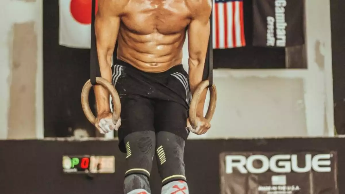
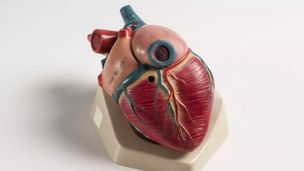
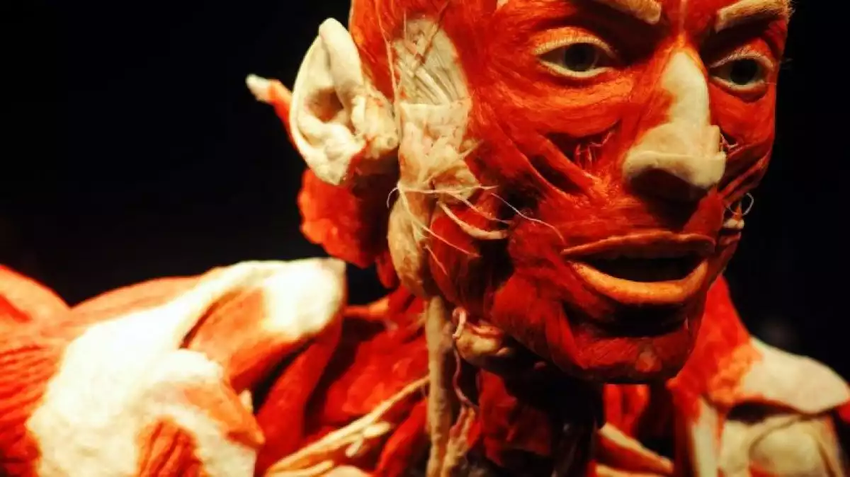
In the human body, the muscles are the set of organs and tissues that make up the musculoskeletal system, which is very important for the movements of our body. In this article, we are going to find out interesting things about human anatomy, focusing on the muscles. We are going to learn what the muscles are and how many muscles the human body has. We will also list the main types, including the ones in the head, neck, thorax, arms, hands, feet and legs.
What are the muscles?
The muscles arethe main tissues of the musculoskeletal system, the apparatus in charge of regulating the movement, the postures and the blood circulation, a function that is performed from muscles in coordination with bones.
They are activated by the action of spinal nerves, whose fibers are of a motor or sensory type, and they are located in different sections of the spinal cord.
Depending on their specific localization, these fibers innervate the skin, the neck or thoracic muscles, or the ones of the limbs. This means they send nerve signals to a specific area of the body, which makes that area take action.
Each of the muscles of the human body is formed by different tissues, which are known as "muscle tissues." It is a network of cells that are in charge of giving structure to the organs.
The main type of tissues that make up human musculature are the striated muscle tissue (skeletal muscle), smooth muscle tissue, and cardiac muscle tissue.
Nutritionally speaking, the substances that have the property of increasing the muscle mass (the volume of the muscles) are proteins. However, to keep ourselves healthy and allow blood and oxygen to flow through them, the consumption of vitamins and minerals is also essential.
How many muscles are in the human body and what are the different types?
As we have mentioned before, in the human body there are three main muscle tissues. So, as a whole, these tissues make up the total musculature.
Voluntary muscles (those of the extremities) are made of striated muscle tissue, also called skeletal muscle tissue. Involuntary muscles (those of the internal organs) are made of smooth muscle. The heart, on the other hand, is made almost entirely of cardiac muscle tissue.
So, the ones that allow movement, that is, those in the head, the thorax and the extremities, are a combination of striated muscle tissues.
It has been calculated that the human body has around 650 muscles of this type. The striated types are divided into two significant parts: one axial, comprising the head, neck, and trunk, and another appendicular, formed by the extremities (arms and legs).
In the following lines, we will see which are the different types of muscles formed by striated tissue that make up the axial part and the appendicular one of the human body, starting by the head and going down to the feet.

Head muscles
The head is the top part of our body. It has one of the vital organs in it: the brain. It is also the center of our senses and one of the first contacts with the world. In this part, we find mainly facial muscles, which we will describe below.
Facial muscles
The top part of our face is composed of a temporal muscle, located on both sides of the head (in the area of the temples); a frontal one, which is in the area of the forehead; and an orbicular muscle (round or circular) of the eyelids, which is the one that surrounds the eyes and allows them to open and close.
On the other hand, in the lower part of our face, there are two muscles called "zygomaticus major." There is one in each cheek, which allows the mobility of most of our face. They help us to vocalize, smile, chew, yawn, among many other things.
Just below this facial muscle, there is another one called masseter. This is the one that helps us elevate the lower jaw.
In the same zone we have another orbicular muscle, but this time it is placed in the lips. This muscle allows us to open and close our mouth.
Neck muscles
The neck is the part of our body that connects the head with the body. The three main neck muscles are the anterior scalene, the platysma, and the sternocleidomastoid.
The anterior scalene muscle gets its name because of the irregular triangle shape it has, and also because of where it is located: at one side of the neck. It fixes the spinal column and facilitates its movement.
The platysma muscle is attached to the skin. This muscle participates in an important way in many facial expression, especially during stressful periods.
Under the platysma, we can find the sternocleidomastoid, which gives movements to the joints of the skull, the vertebrae, and the cervicals. Its activity is essential if we want to flexion the neck and head, as well as for innervating the thorax. Therefore, it is also vital to carry out necessary actions such as breathing.
Thoracic muscles
The thorax is also known as the trunk because it is the part of the body that connects to the neck and the head with the extremities. It is where most of our internal organs are, for instance, the heart or the lungs.
Abdominal muscles
The main muscles in the abdomen are called pectorals. We have a pectoralis major and a minor. The first one is located in the most upper part of the abdomen, it is composed of subcutaneous tissue and is primarily responsible for giving movement to the shoulder.
On the other hand, the pectoralis minor is a thinner, inner muscle and also helps the mobility of the shoulder, although it participates less than the major one. It is also responsible for the movement of the ribs.
Other abdominal muscles are the intercostal muscles, which are between the ribs (they are essential for breathing); and the serratus anterior, in the upper part of the thorax (it allows the mobility of the abdomen and the arms).
In the lower part of this area, we find four: an internal oblique and an external oblique; the rectum of the abdomen; and the transverse of the abdomen. And towards the pelvis, there is the tensor of the fascia lata, which is already part of the legs. All of them are fundamental to allow flexion and abdominal posture.
Back muscles
The back is the back part of the thorax. Its main structure is the vertebral column or spine. The latter is a significant part of the musculoskeletal system because it is in charge of moving the extremities, especially the legs.
To achieve this, this structure is formed by the dorsal muscles. The main muscles from this area are the ones in the back of the neck, which regulate the movement of the neck and the shoulders. These are the occipital muscle, the sternocleidomastoid, the splenium of the head and the trapezium.
The back muscle we find in the upper part of it is the latissimus dorsi, which is the strongest one of the trunk part of the body. A bit higher up, towards the back of the neck, we have the levator scapulae muscle and the rhomboids (a major and a minor).
Towards the shoulders, and in their upper part, there is the supraspinatus muscle; while in the lower part we find the infraspinatus muscle. And at the bottom of this last one, there are the teres (there's also a major and a minor).
In the middle part of the back, there is another important muscle for the movement of the trunk. It is called the erector spinae muscle, and just below it, there are the external intercostal muscles, which are also part of the abdomen.
To finish, in the lower part of the back we have the lumbar triangle, the gluteus medius, and the gluteus maximus.
Shoulder muscles
The shoulder muscles are also known as deltoid muscles. They are around the joints of each shoulder, and they are divided into 3 different regions. Each one of them is in charge of allowing the shoulder to move in different directions.

Cardiac muscle
As we have previously said, the heart is made of a muscle tissue called "cardiac muscle tissue or myocardium." These cells make up more than half the heart.
Because of this tissue, whose innervation is involuntary, our heart can beat. This movement allows the blood to get to the rest of our body.
Hands and arms muscles
The arms are the upper limbs of the human body, and in their most external parts, they have the hands and the fingers, while in their inner part they have the forearm. The part nearest to the neck is called shoulder.
Each arm has the following muscles, whose function is to flex the elbows and allow movement to the forearms.
Biceps brachii
Triceps brachii
Brachioradialis
There are also other muscles that take part in the elbow and the forearm's flexion. If we look at the arms from the front, we can see the following: the brachialis, the flexor carpi radialis, the palmaris longus, flexor carpi ulnaris, and the pronator teres.
Forearms and hands
None of the previously mentioned muscles is part of the forearm, but they help with the movement when we bend the elbow.
Thus, the forearm muscles (if we look at the back of it) and that also allow the movement of the fingers are these: the anconeus muscle, the extensor carpi ulnaris muscle, the extensor carpi radialis brevis muscle, the abdominal external oblique muscle, the flexor carpi ulnaris, the extensor digitorum muscle, and the infraspinatus.
Feet and leg muscles
The leg is divided into different muscles depending on the specific part of this extremity. We can learn the leg muscles by observing them from the front or the back.
Thigh muscles
In the upper part, known as the thigh, there are seven leg muscles (if we focus on the front of it):
Pectineus, which is in charge of moving the thigh in a direction known as adduction (outside to inside).
Adductor longus, that allows the movement of the pelvis above the thigh.
Sartorius, in the human body, is the most extensive muscle. It is responsible for moving both the hip and knees.
Rectus femoris, It is responsible for moving the quadriceps.
Vastus lateralis, like the previous muscle, it allows the movement of the quadriceps.
Vastus medialis, together with the last two muscles, make up the quadriceps.
Biceps femoris, which is located just behind the gluteus and gives strength to the upper body.
On the other hand, if we look at the thigh from behind we find:
Adductor magnus, which is just under the gluteus.
Gracilis, very important to for moving the hips.
Iliotibial band, that gives mobility to the knees.
Semimembranosus, which directs the back
Semitendinosus, that acts both on the back and on the knee.
Shin
The lower part of the leg is commonly known as calf (behind) and shin (front). In the latter there are the following muscles:
Extensor digitorum longus, which is important for the movements of the toes.
Peroneus longus, whose contraction helps rotate the feet.
Tibialis (anterior and posterior), it is the main muscle of the feet, for it allows its flexion.
Calf
At the back of the leg (the calf) there are the following muscles:
Gastrocnemius, also known as calf muscles (they are divided into two), and has the function of flexing the soles of the feet.
Soleus, it is located on the lower part of the legs, and it helps to flex the feet.
Plantaris, which is in a more internal area than the calf and helps to lift the feet and move the knees, although with less involvement.
Achilles tendon, which is an extension of the soleus muscle and the plantaris and also allows the feet to move.
- This article about "The Muscles" was originally published in Spanish in Viviendo La Salud
References
Jarmey, C. (2008) Atlas conciso de los músculos. Editorial Paidotribo: España.
Latarjet, M. & Ruiz, A. (2006). Anatomía humana. Editorial Médica Panamericana: Argentina.
Palastanga, N., Field, D. & Soames, R. (2000). Anatomía y movimiento humano. Estructura y funcionamiento. Editorial Paidotribo: España.
