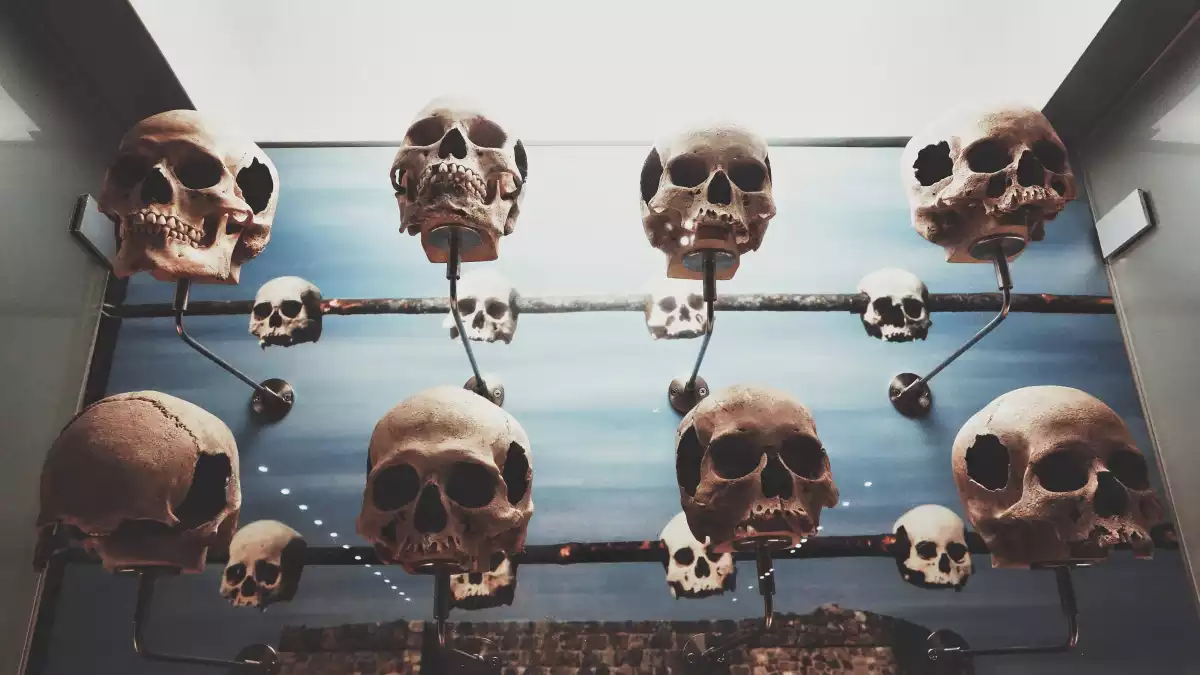
The human skull is a bony structure that contains the brain with its cerebral hemispheres, cerebellum, and nervous system and one of the primary functions of the skull is to protect from injury.
We will examine the different parts of the human skull, the muscles associated with them and their different functions and answer the questions: "How many bones are in the human skull?" and "How thick is the human skull?".
Human skull - Parts and function
Skulls are a typical structure in all vertebrate animals. The human skull is formed with the help of articulations called brain sutures, and the main tasks of the skull are to protect and support the brain and all the nervous systems that are encased in it.
From an anatomical perspective, the skull is divided into two parts: the cranium and the mandible. These are also called neurocranium - braincase, and viscerocranium - facial skeleton.
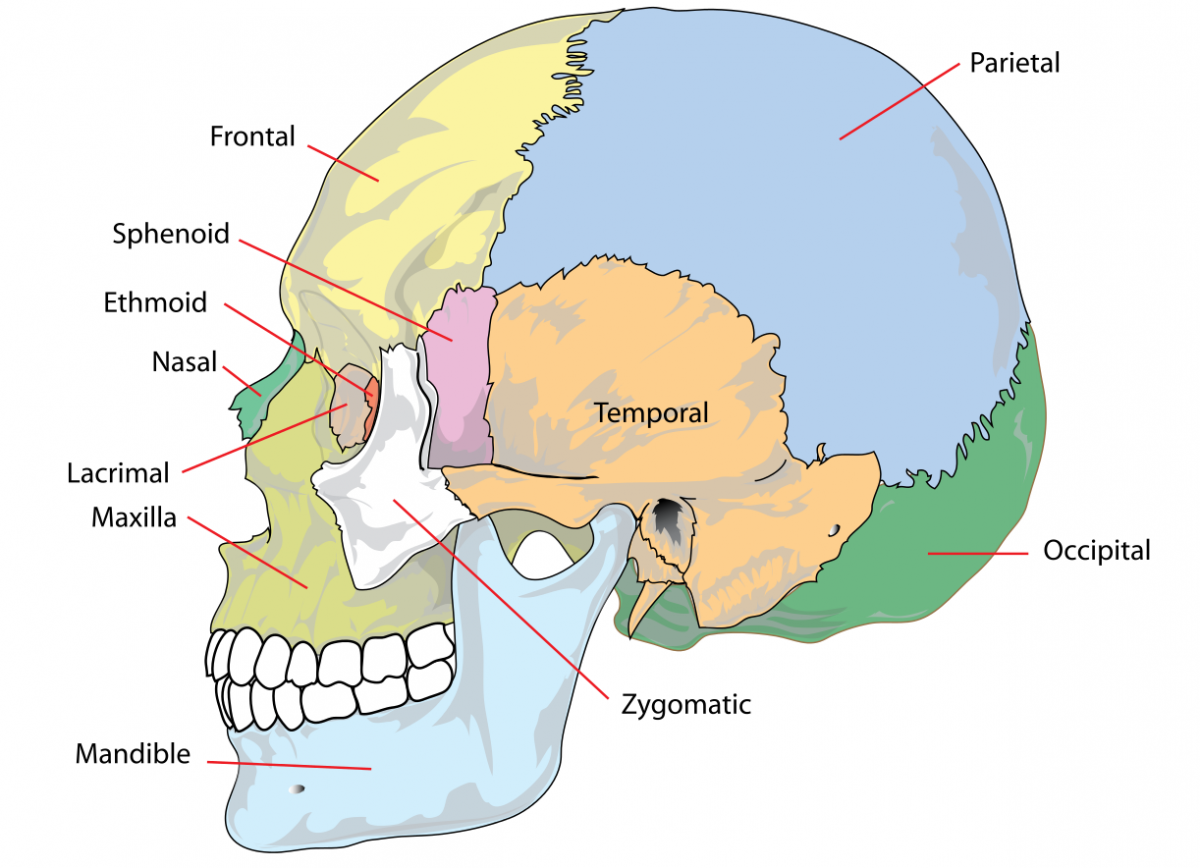
The upper side of the brain includes the frontal bone, the occipital, parietal and temporal bones and together they form the neurocranium.
Another possible division of the human skull is to differentiate between the endocranium - inner part of the skull, and the ectocranium - outer portion of the skull.
Human skull - Bones
The human skull is generally considered to consist of twenty-two bones—eight cranial bones and fourteen facial skeleton bones.
The average thickness of a male skull is 0.25 inches (6.5 millimeters), while the average thickness of a female skull is 0.28 inches (7.1 millimeters).
The neurocranium, also known as the braincase, is the upper and back part of the skull, which forms a protective case around the brain and it contains the following bones:
- two temporal bones
- two parietal bones
- a frontal bone
- an occipital bone
- the sphenoid
- ethmoid bone
On the other hand, the facial skeleton, the lower half of the human skull, is comprised of the mandible, the maxilla, and the rest of the nasal bones.
The human skull bones are connected via a series of suture joints. Sutures form a tight union that prevents movement between the bones. Most sutures are named for the bones they articulate, and the main four brain sutures are the following:
- the coronal suture - joins the frontal bone to the parietal bones
- the sagittal suture - joins the two parietal bones to each other
- the lambdoid suture - joins the parietal bones to the occipital bone
- the squamous suture - joins the parietal bones to the temporal bone

1. Frontal
The frontal bone of the human skull is a bowl-shaped bone in the forehead region of the skull. It also connects to the base of the skull and the ocular orbits.
2. Temporal
The temporal bones are situated at the sides and base of the skull, and lateral to the temporal lobes of the cerebral cortex.
3. Parietal
The parietal bones are two bones in the human skull which, when joined together at a fibrous joint, form the sides and roof of the skull.
4. Occipital
The occipital bone is the bone found at the lower-back area of the cranium. The occipital muscle is cupped like a saucer to accommodate the back part of the brain. Here, we find the foramen magnum which connects the base of the skull with the spinal cord.
5. Ethmoid
The ethmoid bone is a small bone located between the frontal and the sphenoid and separates the skull from the nasal cavity.
6. Sphenoid
The sphenoid bone is an unpaired bone of the neurocranium. It is situated in the middle of the skull towards the front, in front of the temporal bone and the base of the occipital bone.
7. Nasal
The nasal bones are two small rectangular bones; they are placed side by side at the middle and upper part of the face and by their junction, form the bridge of the nose.
8. Vomer
The back portion of the bridge of the nose is formed by the vomer bone which separates the two nostrils.
9. Lacrimal
The lacrimal bone is a small bone of the facial skeleton; it is roughly the size of the little fingernail. It is situated at the front part of the medial wall of the orbit.
10. Zygomatic
In the human skull, the zygomatic bone (cheekbone or malar bone) is a paired irregular bone which articulates with the maxilla, the temporal bone, the sphenoid bone, and the frontal bone.
11. Maxilla
The maxilla bone is the most important bone of the face (in the viscerocranium). It contains the maxillary sinus, a cavity whose walls are lined with a mucous membrane.
12. Mandible
The mandible, lower jaw or jawbone is the largest, strongest and lowest bone in the human face. It forms the lower jaw and holds the lower teeth in place. The mandible sits beneath the maxilla.

Human skull - Facial muscles
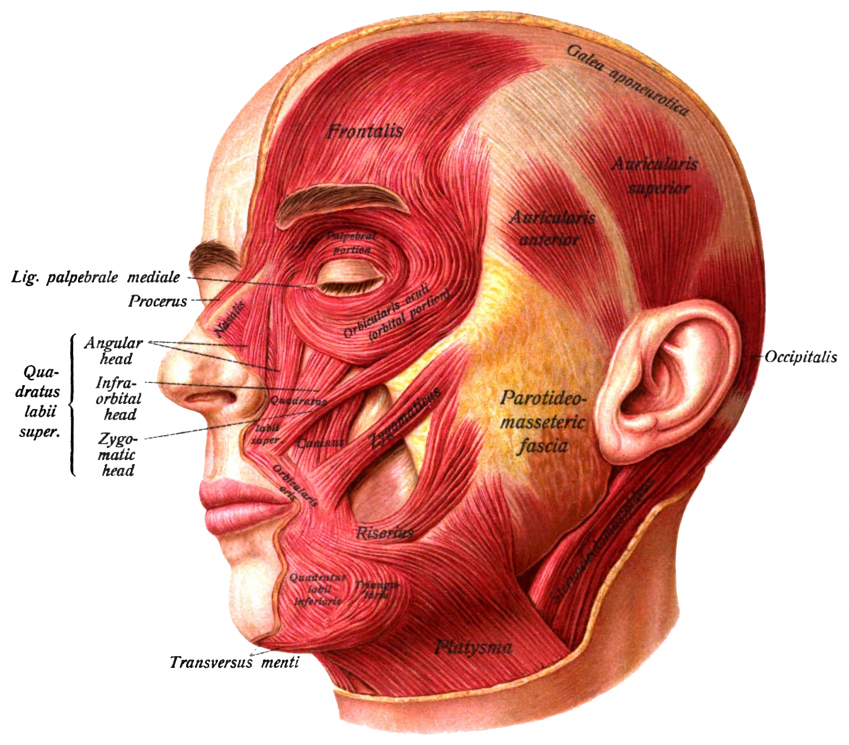
The facial muscles are a group of striated skeletal muscles that control facial expressions and allow us to chew our food. The muscles located on our skull are the following:
Occipitofrontalis muscle - the large muscle of the forehead
Temporalis muscle - a thick muscle that closes the mouth and assists the jaw to move side-to-side to grind up food
Procerus muscle - a small pyramidal slip of tissue in between the eyebrows, helps flare the nostrils and express anger
Nasalis muscle - the muscle responsible for "flaring" of the nostrils
Depressor septi nasi muscle - this muscle arises from the incisive fossa of the maxilla, closes the nasal septum
Orbicularis oculi muscle - muscle in the face that closes the eyelids
Corrugator supercilii muscle - muscle functions to move the eyebrow down and inward toward the nose and inner eye, creates vertical lines or wrinkles
Auricular muscles (anterior, superior and posterior) - the outer ear, external ear, or auris externa is the external portion of the ear, which consists of the auricle (also pinna) and the ear canal. It gathers sound energy and focuses it on the eardrum
Orbicularis Oris muscle - this muscle brings our lips together so we can pucker up for a kiss
Depressor anguli oris muscle - facial muscle associated with frowning, starts from the mandible and inserts into the angle of the mouth.
Risorius - a very thin and delicate muscle that pulls the lips horizontally creating a broad, albeit insincere smile
Zygomaticus major muscle - draws the angle of the mouth superiorly and posteriorly to allow one to smile
Zygomaticus minor muscle - it brings the upper lip backward, upward, and outward and is used in smiling
Levator labii superioris alaeque nasi - this muscle dilates the nostrils and raises the upper lip. It’s often referred to as the ‘Elvis muscle’ in homage to Elvis Presley.
Depressor labii inferioris muscle - this muscle pulls down the bottom lip allowing us to sulk
Levator anguli oris - the happy muscle, making the corners of our mouth turn upwards into a smile
Buccinator muscle - known as the ‘trumpeter muscle,’ the Buccinator’s role is to puff out the cheeks and prevent food from passing to the outer surface of the teeth during chewing
Mentalis - called the ‘pouting muscle,’ contraction of the Mentalis raises and thrusts out the lower lip to make us pout
Check out the original article: Cráneo humano: partes, huesos, músculos y anatomía at viviendolasalud.com
References:
Carlson, B. M. (1999). Human Embryology & Developmental Biology. Mosby.
Rouvière, H. & Delmas, A. (1996). Anatomía humana: descriptiva, topográfica y funcional (9ª Ed.), Tomo I. Masson.
Slater, B. J., Lenton, K. A., Kwan, M. D., Gupta, D. M., Wan, D. C. & Longaker, M. T. (2008). Cranial sutures: a brief review. Plastic and Reconstructive Surgery, 121(4): 170e–8e.
FORENSIC FACIAL RECONSTRUCTION, THE UNIVERSITY OF SHEFFIELD, https://www.futurelearn.com/courses/forensic-facial-reconstruction/0/steps/25674
